Pulmonology focuses on diagnosing and treating conditions related to the lungs and respiratory system. Issues such as airway blockages, fluid accumulation, lung infections, and pleural diseases can significantly affect breathing and overall health, requiring prompt and specialized intervention.
Dr. Ravi Manek provides advanced, minimally invasive, image-guided pulmonology procedures designed to improve lung function and relieve respiratory symptoms. Treatments such as pleural drainage, biopsy, bronchial interventions, and fluid management offer precise results with minimal discomfort.
These procedures ensure quicker recovery, reduced complications, and effective long-term relief. Each treatment plan is customized to address the patient’s specific condition, supporting better breathing, improved mobility, and enhanced respiratory health.
Haemoptysis embolisation is a minimally invasive procedure which deliberately blocks the bleeding vessel, such as the bronchial arteries or pulmonary veins.
The interventional radiologist will insert a 2-3 mm tube into your groin and will guide it under imaging to the affected blood vessel. Small resin particles (microparticles) or small metal spirals (coils) will be inserted into the bleeding vessel or vessels. This causes the vessel or vessels to become blocked and so stops the bleeding.
There are two main reasons why it is important to treat haemoptysis. If too much blood is lost, the patient may go into shock, which is life-threatening. There is also the risk of the patient inhaling the blood: if the patient breathes in too much blood, they may drown
Empyema is defined by purulent fluid collection in the pleural space, which is most commonly caused by pneumonia. A lung abscess, on the other hand, is a parenchymal necrosis with confined cavitation that results from a pulmonary infection.
Some patients may need both antibiotics and a chest drain. A chest drain is a flexible plastic tube that’s inserted through the chest wall and into the affected area to drain it of fluid. The area where the tube is inserted is numbed, and the patient may also be given a light sedative before having the drain inserted.
Radiofrequency ablation (RFA) and microwave ablation (MWA) are treatments that use image guidance to place a needle through the skin into a tumor within the chest.
In RFA, high-frequency electrical currents are passed through an electrode, creating a small region of heat.
In MWA, microwaves are created from the needle to create a small region of heat.
The heat destroys the lung cancer cells.
RFA and MWA may be used to treat lung tumors or provide palliative care.
They are effective treatment options for patients who might have difficulty with surgery and for those for whom surgery is not an option due to the spread of a tumor to the lungs from the primary tumor or cancers outside the chest.
RFA can usually be administered as an outpatient procedure, though may at times require a brief hospital stay.
Pleural effusion, sometimes referred to as “water on the lungs,” is the build-up of excess fluid between the layers of the pleura outside the lungs. The pleura are thin membranes that line the lungs and the inside of the chest cavity and act to lubricate and facilitate breathing.
A pleural aspiration is a procedure where a small needle or tube is inserted into the space between the lung and chest wall to remove fluid that has accumulated around the lung. This space is called the pleural space.
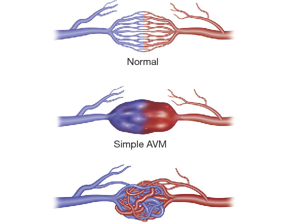
Pulmonary arteriovenous malformation (PAVM) is a rare anomalies of the lung that affects blood flow between the heart and the lungs.
Affected people have an abnormal connection between the pulmonary vein (carries blood from the lungs to the heart) and pulmonary artery (carries blood from the heart to the lungs).
Some people will have more than one PAVM; in more than half of patients with multiple PAVM this is associated with a condition called Hereditary Haemorrhagic Telangectasia.
Pulmonary arteriovenous malformations are usually congenital.
Most patients are asymptomatic.
The aims of treatment of PAVM are to block the PAVM and reduce the risk of stroke, heart failure and improve breathlessness.
Endovascular therapy is a minimally invasive outpatient based treatment wherein the feeding artery to the PAVM is occluded with coils or plugs or a combination of both.
This endovascular treatment associated with minimal morbidity and no mortality.
It is a condition in which one or more arteries in the lungs become blocked by a blood clot.
Most times, a pulmonary embolism is caused by blood clots that travel from the legs, or rarely, other parts of the body (deep vein thrombosis or DVT).
Symptoms include shortness of breath, chest pain and cough.
It is treated by Catheter-directed pulmonary thrombolysis
Catheter-directed pulmonary thrombolysis treats vascular blockages and improves blood flow of pulmonary circulation by dissolving abnormal blood clots.
A blood clot, or thrombus, can block off blood supply to lungs and cause serious damage.
Image-guided, minimally invasive procedures such as thrombolysis are performed by a specially trained interventional radiologist in an interventional radiology suite.
To treat the clot with medication, the catheter is left in place and connected to a special machine that delivers the medication at a precise rate.
Clot-dissolving medications are delivered through the catheter over several hours to a few days. It may take up to 72 hours for the clot to dissolve (although most clots dissolve within 24 hours), during which time patient will be monitored closely by the medical staff.
When the procedure is complete, the catheter is removed and pressure is applied to stop any bleeding. No stitches are visible on the skin. The tiny opening in the skin is covered with a dressing.
Catheter-directed thrombolysis can greatly improve blood flow and reduce or eliminate the related symptoms and effects without the need for more invasive surgery.
Thrombolysis is a safe, highly effective way of re-establishing circulation blocked by a clot.
Thrombolysis is less invasive than conventional open surgery to remove clots and the hospital stay is relatively brief. Blood loss is less than with traditional surgical treatment and there is no obvious surgical incision.
No surgical incision is necessary—only a small nick in the skin that does not need stitches.